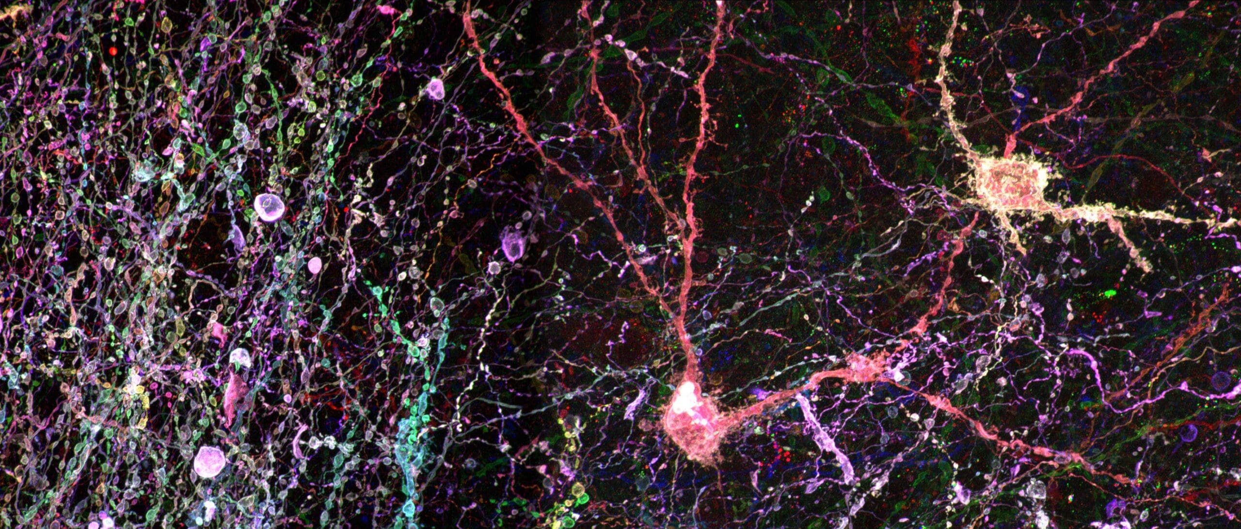
My laboratory develops the wet lab and dry lab technologies needed to map molecular connectomes of the Drosophila and mouse brain, and then uses those maps to determine how the molecular and connectivity identity of a neural circuit shapes somatosensory processing and behavior. Data science is not a downstream step in this program. It drives our experimental design, because a single expanded and multiplexed brain sample now yields terabyte- to petabyte-scale volumetric image data that cannot be annotated by hand.
To generate these data, we combine combinatorial genetic labeling, sample expansion, whole organ processing, and multi-round spatial multi-omics with high-throughput volumetric microscopes that we design in-house. Each choice is made to raise the information content per voxel, because the downstream problem is fundamentally one of separating many overlapping molecular and structural channels.
Our computational program then turns those voxels into biology. We develop deep learning and computer vision methods for dense neuron tracing, cell instance segmentation, and structure annotation in multispectral images. We build probabilistic frameworks that decode combinatorial fluorescent barcodes into cell identity and lineage assignments with quantified confidence, and informatics approaches that correlate transcriptional identity, morphology, connectivity, and physiology within the same neurons. We maintain the resulting tools as open source software for the neuroscience community. We also address the infrastructure problem directly, building browser-based platforms for storing, visualizing, and analyzing petabyte-scale 3D images in high-performance computing environments, along with compression methods that preserve downstream analysis fidelity rather than perceptual quality alone.
I am eager to work with the MIDAS community on foundation models for biological structure annotation, on benchmark datasets and evaluation standards for dense 3D neuroanatomy, and on the data management problems that increasingly constrain what modern microscopy can deliver.
Please describe one or two of your most interesting projects.
Reconstructing the molecular connectome of the Drosophila and mouse brain. A wiring diagram alone cannot explain how a circuit computes, and a molecular census alone cannot say who talks to whom. This project fuses the two into one atlas in which every reconstructed neuron carries its morphology, its synaptic partners at synapse level precision, and its molecular identity. We generate the raw data with in-house sample expansion chemistry, multiplexed spatial omics, and custom high-throughput volumetric microscopes, then reconstruct it using deep learning tracing and segmentation models, probabilistic decoding of combinatorial fluorescent barcodes, and registration methods that place molecular measurements back onto reconstructed arbors. A single brain yields petabyte-scale imagery, so compression, storage, and analysis infrastructure are as much a part of the project as the microscopy itself.
How the brain perceives and modulates facial somatosensory information, especially pain. Orofacial sensation is carried by trigeminal circuits that are molecularly diverse and drive behaviors we can measure precisely, which makes them an ideal testbed for the atlas approach. We ask which molecularly defined sensory and central neuron subtypes carry innocuous touch versus noxious input, how those signals are gated and modulated, and how that gating shifts in injury and chronic pain states. Computationally, this means combining single cell and spatial transcriptomic profiling with circuit tracing and activity recording, applying machine learning to annotate behavior from video, and building informatics tools that correlate gene expression, connectivity, and behavioral phenotype within the same animals. Recent work with U-M collaborators traced a touch-guided circuit that drives gnawing to maintain dental alignment, linking molecular identity, wiring, and behavior end to end.
What makes you excited about your data science and AI research?
For most of my career, the limiting factor in neuroscience was how much data we could generate. That has inverted. My lab can now expand, label, and image a whole brain at a resolution that resolves individual synapses along with the molecules that define them, and the bottleneck has moved entirely to interpretation. One sample produces more image data than a trained anatomist could annotate in a lifetime. That inversion is what excites me, because it means the questions we can ask are now set by our algorithms rather than by our microscopes.
What I find most rewarding is that the computational problems are genuinely hard and genuinely general. Tracing thousands of entangled, faintly labeled processes through a noisy 3D volume, decoding combinatorial barcodes into confident cell identity calls, and correlating gene expression with wiring and behavior in the same animal are problems that push machine learning methods rather than simply applying them. The methods we build for brain tissue transfer readily to other dense biological imaging domains.
I also enjoy the collaborations this work demands. Progress here requires chemists, optical engineers, computer vision researchers, and neuroscientists working on the same problem at the same time, and the most interesting ideas in my lab have almost always come from the boundary between two of those fields rather than the middle of any one of them.