Search
Affiliation Type
Methodologies
Applications

Keith Aaronson, MD
Bertram Pitt M.D. Collegiate Professor of Internal Medicine - Cardiovascular Medicine, Michigan Medical School

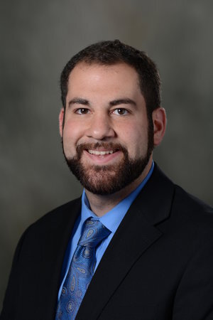
Egide Abahuje
Assistant Professor of Cardiac Surgery, Michigan Medical School

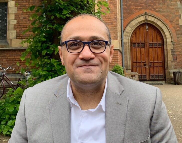
Ahmed Abdel-Latif
Clinical Professorof Internal Medicine - Cardiology, Program Director of Internal Medicine, Michigan Medical School
Yasser Aboelkassem
Assistant Professor of Engineering, College of Innovation and Technology, University of Michigan - Flint

Mohamed Abouelenien
Associate Professor of Computer and Information Science, College of Engineering and Computer Science, The University of Michigan - Dearborn
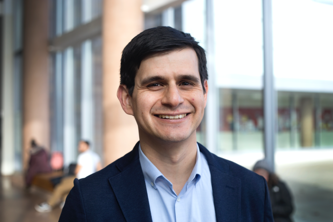
Andrew J. Admon, MD, MPH, MSc
Assistant Professor of Internal Medicine, Michigan Medical School
Assistant Professor of Epidemiology, School of Public Health

Michelle Aebersold
Clinical Professor of Systems, Populations, and Leadership and Academic Program Director, School of Nursing
Clinical Associate Professor of Information, School of Information

Mahesh Agarwal
Chair and Associate Professor of Mathematics and Statistics, College of Arts, Sciences and Letters, The University of Michigan-Dearborn
Carlos Aguilar
Associate Professor of Biomedical Engineering, College of Engineering and Michigan Medical School