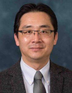
My research focuses on novel biomedical imaging and treatment technologies, especially those involving light and ultrasound. I have extensive experience in medical system development, laser-tissue interactions, ultrasound tissue characterization, and adaptation of novel technologies to preclinical and clinical settings. A major part of my research is the development and clinical applications of photoacoustic imaging technology. By working on small-animal models and human patients, I have been seeking for clinical applications of this exciting technology to inflammatory arthritis, cancer, inflammatory bowel disease, eye diseases, osteoporosis, and brain disorders. Besides photoacoustic imaging, I am also interested in development of other medical imaging and treatment technologies, such as ionizing radiation induced acoustic imaging (iRAI) and photo-mediated ultrasound therapy (PUT). As the PI or co-Investigator of several NIH, NSF and DoD-funded research, I have successfully administered the projects, collaborated with other researchers, and produced high-quality publications. My contribution to biomedical optics and ultrasound up to now including over 160 peer-reviewed journal papers is a solid evidence of my creativity and ability to surmount the challenges in this field. I received the Sontag Foundation Fellow of the Arthritis National Research Foundation in 2005, the Distinguished Investigator Award of the Academy of Radiology Research in 2013, and was elected as the fellow of AIMBE in 2020 and the fellow of SPIE in 2022.
What is your most interesting project?
Automated photoacoustic imaging of inflammatory arthritis: Our research has demonstrated the unique capability of photoacoustic imaging (PAI) in diagnosis and treatment monitoring of inflammatory arthritis. The new physiological and molecular biomarkers of synovitis presented by PAI can help in characterizing disease onset, progression, and response to therapy. Based on the endogenous optical contrast, PAI is extremely sensitive to the changes in hemodynamic properties in inflammatory joint tissues (e.g. enhanced flow and hypoxia). We are now conducting a preclinical research on patients affected by rheumatoid arthritis. The initial findings from this patient study are promising and suggest that the new optical contrast and physiological information introduced by PAI could greatly enhance the sensitivity and accuracy of diagnostic imaging and treatment monitoring of arthritis. Aiming at clinical translation, we are currently developing a point-of-care PAI and ultrasound dual-modality imaging system which is fully automated when powered by a robot and AI technologies.
Ionizing radiation acoustic imaging (iRAI) for personalized radiation therapy: iRAI, as a brand-new imaging technology relying on the detection of radiation-induced acoustic waves, allows online monitoring of radiation’s interactions with tissues during radiation therapy, providing real-time, adaptive feedback for cancer treatments. We are developing an iRAI volumetric imaging system that enables mapping of the three-dimensional (3D) radiation dose distribution in a complex clinical radiotherapy treatment. The feasibility of imaging temporal 3D dose accumulation was first validated in studies on phantoms and animal models. Then, real-time visualization of the 3D radiation dose delivered to a patient with liver metastases was accomplished with a clinical linear accelerator. These studies demonstrate the great potential of iRAI to monitor and quantify the 3D radiation dose deposition during treatment, potentially improving radiotherapy treatment efficacy using real-time adaptive treatment.
Describe your research journey.
2005 – 2007 Research Investigator, Department of Radiology, University of Michigan
2007 – 2008 Research Assistant Professor, Department of Radiology, University of Michigan
2008 – 2012 Assistant Professor, Department of Radiology, University of Michigan Medical School
2012 – 2014 Associate Professor, Department of Radiology, University of Michigan
2015 – 2018 Associate Professor, Department of Biomedical Engineering, University of Michigan
2018 – 2022 Professor, Department of Biomedical Engineering, University of Michigan
2022 – Now Jonathan Rubin Collegiate Professor of Biomedical Engineering, University of Michigan
What is the most significant scientific contribution you would like to make?
Develop and translate state-of-the-art medical imaging and treatment technologies.
What makes you excited about your data science and AI research?
Date science and AI is super important in developing state-of-the-art medical imaging and treatment technologies, especially for achieving personalized diagnosis and treatment ensuring largely improved patient outcome. As mentioned in the above, the automated imaging system for rheumatology/radiology clinic for arthritis imaging would be strongly powered by AI, which is crucial to achieve our goal of a “smart” ultrasound imaging platform.
